|
Natural
Growth Factors Affect on the Vascular |
|
I.
Mikadze, G. Abzianidze Key words: Growth factors, vascular prosthesis, myointimal hyperplasia, endothelium, neointima, angiogenesis. |
|
Tissue
GFs have great significance for angiogenesis, process of healing of
vascular prostheses and myointimal hyperplasia, that leads to stenoses
of implanted prostheses and causes their thrombing [2-5]. Mechanism
of action of different GFs is still unknown. However, it’s possible to
determine types of cells, affected by different GFs. Among lots of GFs,
collaborating in regulation of angiogenesis, the most important are
representatives of fibroblast GF family (FGF-1, FGF-2, FGF-5) and
vascular endothelial GF (VEGF), transforming GF b (TGF-b).
These mitogens are combined by their ability to stimulate growth of cells
of mesenchymal row and in particular of endothelium.
Affect
of natural GFs on vascular prostheses healing has not been completely
investigated yet. Successful use of FGF and VEGF for angiogenesis
stimulation in experiment and in clinic testifies the necessity of
research in this area [7, 8]. It
is still unclear, if surplus concentration of GFs in periprostheses
tissues is the initial mechanism for myointimal hyperplasia, how GFs
affect the formation of vascular wall prostheses grafting, and if it is
possible to improve endothelisation of implanted vascular prosthesis,
stimulating growth of mesenchimal cells by polypeptide GFs.
To
answer these questions we investigated healing of arterial substitutions,
treating them with brain obtained from artiodactyls tissue GF. Materials
and Methods
The growth
factor (GF) was obtained from the bovine brain according to Magiac T.
(1982) method. The analysis of the obtained material was made on
polyacrylamide gel plates, comprising 10%-ed division and 3%-ed concentration gel in presence of dodecyl
sulphate of natrium. affect of different concentrations of GF and heparin on the growth of endothelial cells in proportion 2:1 was observed in culture. The following concentrations of GF were investigated: 100; 200; 400; 600 mg/ml of medium. On the gelatin coated Petri dish with diameter of 40 mm endothelial cells of umbilical cord vein were seeded in the amount of 30 ´ 103 - 35 ´ 103 cells per dish that were harvested from monolayer culture and were cultivated in similar conditions. The medium was changed every other day. The cultures of endothelial cells were trypsinized and counted in 24, 48, 72 hours. To
determine the proliferative pool, all the cells of culture were counted,
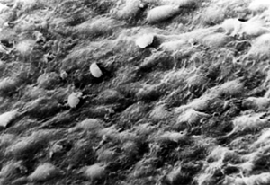
including the dead ones after the treatment of trypsin. To study the GFs affect on the vascular prosthesis and arterial pitch healing we used PTFE ("GORE", “IMPRA”), fluorolavsan prostheses, xenoprostheses, and human durra mater pitches. The treatment of prostheses by the GF was made by the original method. Prostheses passed through alcohol battery of 30%-50%-90%-100% for 5 min in each concentration. Then they were aired for 2-3 min and placed in heparinized plasma. Plasma was taken from the dog prepared for operation 1-2 days earlier. GF was added in autoplasma in 4 different concentrations: 10.0 mg % – 5.0 mg % - 0.5 mg % - 0.05 mg %. Antibiotics, prednizolon were added to the plasma in quantity that is used for cultures of tissues and cells. The redundant heparin effect was removed by supplement natrii protaminsulfurici after the prostheses had been placed in the prepared plasma. The prostheses remained there at 370 C for 2 hours and then kept there at 40 C. Next day the graft was implanted. Experimental animals were divided into 4 groups depending on increasing GF concentration in solution: in the first group of animals, 21 experiments, prostheses were treated with the solution of autoplasma, that had 10 mg% GF (0.05 mg/cm 2 of prosthesis); in the second group of animals, 21 experiments, prostheses were treated by the solution of autoplasma that had 5mg% GF (0.025mg/cm 2 of prosthesis); in the third group of animals, 113 experiments, GF was added to autoplasma in quantity of 0.5 mg % (0.0025 mg/cm 2 of prosthesis); in the fourth group of animals, 94 experiments, GF was added to autoplasma in quantity of 0.05 mg % (0.00025 mg/cm2 of prosthesis). Operations of grafting on the carotids (54), femoral arteries (48), infrarenal part of abdominal aorta (174), torakoabdominal shunting (16) were caried out. All types of pitches were treated by GF in amount of 0.0025mg/cm2 (36 experiments), and0.00025 mg/sm2 of grafts (62 experiments). The sizes of pitches in the carotids and femoralis position were 3x5 and 5x12 mm in the aorta abdominalis position. The pitches were implanted in abdominal aorta, femoralis and carotid arteries of mongrels weighing15-20 kg. Time of observation - up to 6 months. Results and Discussion Obtained GF comprises no less than 13 albuminous components, with molecular mass in measures of 20×103-70×103 Da. The number of endothelial cells in culture with the use of mitogen in concentration 100 mg/ml in 24 hours was 40.4±3.3 ´ 103 doubled in 48 hours and became 65.7±3.4 ´ 103 (р< 0.05). The rise of GF concentration to 200 mg/ml leads to increasing of the cells quantity twice as much already in 24 hours (60.3±1.8 ´ 103). Tempos of growth are kept and the quantity of cells in 48 hours is 100.2±9.2´103 and in 72 hours - 240.2±2.0 ´ 103. The number of cells with the use of mitogen in concentration of 400 mg/ml in 24 hours of cultivating was 55.4±7.4 ´ 103 and authentically didn’t differ from the number of cells in 24 hours with GF used in concentration of 200 mg/ml. Using this concentration of mitogen in culture, cells keep their tempos of growth. The number of cells in 48 hours of cultivating was 110.4±10.8 ´ 103 and in 72 hours - 230.1±4.0 ´103. The rise of concentration of GF to 600 mg/ml didn’t increase the number of cells. The number of cells in 48 and 72 hours in comparison with the use of mitogen in concentration of 400 mg/ml decreased and became 85.7±6.6 ´ 103 and 160.0±2.0 ´ 103 against 110.4±10.8 ´ 103 and 230.1±4.0 ´ 103 agreeably. The most effective concentration of GF is 200 mg/ml in combination with 100 mg/ml of heparin. The use of this concentration of GF and heparin allows to obtain quickly the proliferating culture of endothelial cells within 24 hours of PD. At least 15 passages keep the main characteristics of endothelial cells; they formed the monolayer of cells with the picture “cobblestone”, they have character of contact inhibition of growth, the borders of the cells are stained with silver nitrate, are stained with factor VlllR coagulation of blood, synthesize angiotensin-converting enzyme in quantity of 3-6 nmol on 106 cells in confluent monolayer [1]. Our investigation shows that GF obtained from calf’s brain has mitogen activity to endothelial cells. First and second groups of animals (42 experiments) The prostheses were treated with solution of autoplasma containing 10 mg%(0.05 mg/sm2 of prosthesis); and 5 mg% of GF (0.025 mg/sm2 of prosthesis). In these groups beginning from the first day the prostheses trombosed for the first 2 weeks after implantation. Using prostheses from PTFE and fluorolavsan in first days after implantation the forming of neointima with the thickness much superior than the thickness of prosthesis was noted . In this case neointima is parietal thrombus on different levels of organization. Third group of animals (113 experiments) GF was added into autoplasma in amount of 0.5 mg% (0.0025 mg/sm2 of prosthesis). Neovascularisation of prostheses from PTFE and fluorolavsan on the 7-10 day are noted with well formed penetrated vasa vasorum neoadventitia, with dense connection to surface of the prosthesis. The thickness of prosthesis is penetrated with cells mesenchyme row. Investigation with scanning electro microscopy shows, that in two weeks in position of abdominal aorta side of anastomosis is completely covered with solid cell layer, that has identical construction in comparison with the cell layer of aorta. Prostheses in position of carotid and femoral arteries thormbosed during first week after implantation. All experimental prostheses in 4 – 6 weeks after implantation have good cellular organization of neointima and endothelial coverage of luminal surface (Fig.1). Fig. 1. Prosthesis PTFE treated by GF in a month after implantation. SEM. Magnification ×600
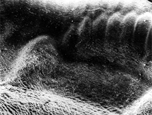
Fourth group of animals (136 experiments) GFs was added in autoplasma in quantity of 0.05 mg%(0.00025mg/sm2 of prosthesis). Patency of prostheses was 90 % during 6 weeks after implantation. In first two weeks anastomoses of prostheses are smooth, shining, color isn’t different from the color of intima of grafted vessel in 1.5-2 sm from anastomosis(2). The narrowing of the prosthesis in the carotid and femoral arteries position, where diameter of prosthesis was 4mm wasn’t observed. Interstice of prosthesis are filled with fibroblasts, smooth-muscled cells, which are over intraluminal surface of prosthesis, where neointima is already formed. Fig. 2. Anastomosis of fluorolavsan prosthesis, 2 weeks after implantation.
SEM. Magnification ×100. The prosthesis is covered by thin thrombus free neointima. Endothelium of aorta fluently crosses the surface of the prosthesis. Anastomoses of prostheses are without any sign of hyperplasia. Formed on the place of wedge-shaped thrombus neointima is thin, covered with endothelial cells, that increase fom the side of artery. Rugosity of surface of artery covers the surface of prosthesis. Endothelium and smooth-muscled cells proliferate from cut ends of artery and form new intima. It is interesting to note, that if endothelial surface hasn’t the whole blast structure, hyperplasia of liable tissue is not observed.(Fig.3). Luminal surface in area of anastomosis is covered by non homogeneous layer of cells that continues proliferating. The area of wedge-shaped thrombus is figured by effuse tissue. The growth of cells in this area possibly leads to stenosis of anastomosis.
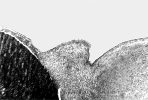
Fig. 3. Anastamosis of controlled PTFE prosthesis in 30 days after implantation. (Hematoxylin –eosin, magnification ×100).
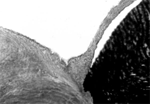
Hyperplasia of neointima is noted. In contrast to control groups, anastomoses of PTFE prosthesis treated with GF don’t have features of hyperplasia. Organized on the place of wedge-shaped thrombus neointima is thin and is covered with solid layer of endothelial cells, growing from the side of artery. Endothelial monolayer has solid structure of monolayer of cells, that smoothly continues from side of grafting artery. Rugosity of surface of artery covers the surface of prosthesis (Fig. 4.). Fig. 4. Anastomosis of PTFE prosthesis treated with GF in 30 days after implantation.
(Picrofuxine on Van-Gizon, magnification ×100).
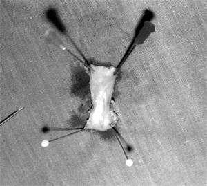
Central part of prostheses has whitish colour, solitary punctate impositions of red color on its background are noted. celled organization of neointima in the centre of prosthesis is observed. Cells of oblong form are directed to blood flow and compile solid monolayer. Fluorolavsan prostheses remain permeable during the investigational period of 180 days, in 100% of cases. Beginning from the first days after implantation neoangiogenesis continues to form consequently. Already in a month, intraluminal surface of prostheses is covered by organized neointima endothelial monolayer. Arterial pitches investigation. In both groups of animals for arterial pitch investigation in two weeks after implantation the wall of PTFE and of durra mater pitch was ingrowthed with mesenchymal cells. In two weeks intraluminal surface was covered with endothelial monolayers. There are no indications of neointima hyperplasia and it is so later after implantation. In a month intraluminal surface of pitch practically doesn’t differ from surface of artery. It has the same color, structure, relief and form (Fig. 5).
Fig.
5. Macropreparation of abdominal aorta of dogs with implantation of patch
from PTFE treated with GF. 2 months after implantation. There
is an opinion, that GFs activate smooth-muscled cells, that begin to
proliferate and migrate from media to intima. For a long time
proliferation of smooth-muscled cells was regarded to be the main process
in evolution of myointimal hyperplasia.
While
endothelium of vessels synthesizes GFs and their inhibitors, that are able
to suppress proliferation of smooth-muscled cells, the forming of vascular
wall depends on quickness and completeness of endothelisation process. We
can assume, that early formation of endothelium on intraluminal surface
stops growth of smoothmuscled cells and prevents from evolution of
hyperplasia. The
absence of hyperplasia of neointima both in area of anastomoses and in the
centre of prosthesis proves that the increase of GF contents in tissues is
not the main mechanism of development of myointimal hyperplasia. GF
use in artery grafting in experiment allows to reach early endothelisation
of vascular substitute luminal surface.
|
References
1.
Abzianidze G., Mikadze I., Moldobaeva A., et al (1989)
Endothelization of Vascular Prostheses by Cells Cultured in Human
Endothelial. Khirurgia. 6, 124-128.
(in Russian). 2. Clowes A.W., Kirkman Th,R., Reidy M. (1986). Mechanisms of Arterial Graft Healing. Americ J Patol. 123, 2, 220-230. 3.
Clowes A.W., Zacharias R.K., Kirkman Th,R. (1987) Early endothelial
coverage of Synthetic Arterial Grafts: Porosity Revisited. Amer. J. Surg. 153,
5, 501 -504. 4.
Golden M.A., Tina Au Y.P., Kenagy R.D., Clowes A.W. (1990). Growth
factor gene expression by intimal cells in healing PTFE grafts. J Vasc Surg. 11, 4, 580-585. 5.
Jarrell B.E., Williams S.K., Hoch J.R., Carabasi R.A. (1987).
Perspectives in vascular surgery-biocompatible vascular surfases:
The past and future role of endothelial cells. Bull. N.Y. Acad. Med. 63,
2, 156-157. 6.
Maciag T., Hoover G.A., Weinstein R. (1982) High and low Molecular
weight form of endothelial cell growth factor. J. Biological Chemistry. 257,
10, 5333-5336. 7. Powell R, Carruth J., Basson M., et al. (1996), Matrix-specific
effect of endothelial control of smooth muscle cell migration. J Vasc Surg.
24, 1, 51-57. 8. Sterpetti A., Lepidi S, Cucina A, et al. (1996). Growth factor
production after polytetrafluoroethylene and vein arterial grafting: An
experimental study. J. Vasc Surg. 23, 3, 453-460.
|